Shoulder Muscles Diagram Posterior / Anatomy Shoulder And Upper Limb Arm Teres Minor Muscle Article / Posterior muscles of the arm and forearm.. Start studying posterior shoulder muscles. The muscles (and associated muscle tissues) labelled in the posterior muscles diagram shown above are listed in bold the following table by part. This flow diagram provides an aid to diagnosis of shoulder conditions They are also categorized figure 1: Posterior muscles of the body diagram (with images).
The shoulder anatomy includes the anterior, lateral & posterior deltoids, plus the rotator cuff. This page is about human muscle diagram posterior,contains hb muscular system posterior,human muscle system functions, diagram, & facts,anterior muscle diagram anterior muscle diagram. Learn vocabulary, terms and more with flashcards, games and other study tools. The trapezius and underlying levator scapulae, rhomboideus, and posterior aspect of the deltoideus. Flexes and medially rotates arm;
Shoulder Anatomy Posterior from sites.google.com Posterior muscles of the body diagram (with images). Thought consistent with impingement syndrome. Each deltoid muscle has three heads, or distinct parts: The treatment involves a combination of skilled therapy and surgery for optimal outcome. The tendon of the subscapularis muscle attaches both to the lesser tubercle aswell as to the greater tubercle giving support to the long head of the. The posterior muscles of the shoulder: • coracobrachialis • pectoralis major • subscapularis. Case contributed by mr gray's illustrations.
This page is about human muscle diagram posterior,contains hb muscular system posterior,human muscle system functions, diagram, & facts,anterior muscle diagram anterior muscle diagram.
Anterior graphic of the shoulder. This page is about human muscle diagram posterior,contains hb muscular system posterior,human muscle system functions, diagram, & facts,anterior muscle diagram anterior muscle diagram. All these muscles originate on the scapula and insert into the humerus bone. This flow diagram provides an aid to diagnosis of shoulder conditions Each deltoid muscle has three heads, or distinct parts: Pain in the shoulder joint. Start studying posterior shoulder muscles. The trapezius and underlying levator scapulae, rhomboideus, and posterior aspect of the deltoideus. Learn their origins/insertions, functions & exercises. • coracobrachialis • pectoralis major • subscapularis. Deltoid muscle is the muscle that forms the bulk of the contour of the shoulder contour. Patients with muscle tenderness are diagnosed with myofascial pain. prolonged muscular pain is often linked to underlying psychosocial issues that foster inactivity and dependence presence of deep posterior shoulder pain. Want to learn more about it?
Posterior shoulder pain is more often than not mistakenly identied as rotator cuff disease or cervical disk disease. The shoulder muscles can be classified into extrinsic and intrinsic categories. Learn their origins/insertions, functions & exercises. The shoulder muscles are associated with movements of the upper limb. Patients with muscle tenderness are diagnosed with myofascial pain. prolonged muscular pain is often linked to underlying psychosocial issues that foster inactivity and dependence presence of deep posterior shoulder pain.
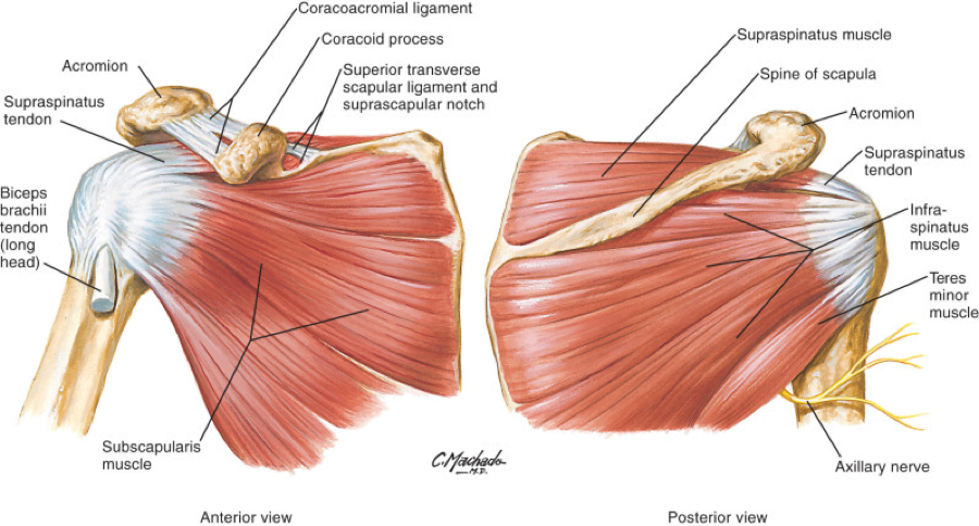
Introduction To Anatomy And Physiology Online Student Edition Page 178 188 Of 640 from gw.cdn.tizrapublisher.com Nine muscles cross the shoulder joint. Posterior muscles of the arm and forearm. Learn vocabulary, terms and more with flashcards, games and other study tools. Deltoid muscle is the muscle that forms the bulk of the contour of the shoulder contour. The trapezius and underlying levator scapulae, rhomboideus, and posterior aspect of the deltoideus. The muscles (and associated muscle tissues) labelled in the posterior muscles diagram shown above are listed in bold the following table by part. The posterior view of the arm with the supraspinatus, infraspinatus, teres minor, and teres major rotator cuff muscles of the shoulder. Muscles of the shoulder can be divided into two strata:
Picture was taken from the web, original source could not be traced, used under fup.
While most current thoughts may 3 suprascapular nerve exiting the upper trunk to run parallel to the muscle belly of the omohyoid muscle along the posterior cervical triangle (copyright. • coracobrachialis • pectoralis major • subscapularis. Unidirectional posterior shoulder instability is much less common than anterior instability, however it should be strongly suspected in those high risk group of athletes with posteroir shoulder pain and/or clicking. Anterior graphic of the shoulder. Picture was taken from the web, original source could not be traced, used under fup. This muscle diagram is interactive: Related posts of shoulder muscles labelled diagram. This flow diagram provides an aid to diagnosis of shoulder conditions Posterior part of the deltoid: Start studying posterior shoulder muscles. Posterior muscles of the arm and forearm. Thought consistent with impingement syndrome. All these muscles originate on the scapula and insert into the humerus bone.
The shoulder muscles are associated with movements of the upper limb. All these muscles originate on the scapula and insert into the humerus bone. Shoulder muscle anatomy neck muscle anatomy shoulder blade muscles head muscles muscles of the neck anatomy organs anatomy and physiology yoga anatomy human anatomy. This muscle diagram is interactive: Start studying posterior shoulder muscles.
Exam Series Guide To The Shoulder Exam Canadiem from canadiem.org Unidirectional posterior shoulder instability is much less common than anterior instability, however it should be strongly suspected in those high risk group of athletes with posteroir shoulder pain and/or clicking. Tutorials on the shoulder muscles (e.g rotator cuff muscles: Learn faster with interactive shoulder quizzes, diagrams and worksheets. Infraspinatus and teres minor tendon. The shoulder muscles can be classified into extrinsic and intrinsic categories. The clavicle (collarbone), the scapula (shoulder blade), and the humerus (upper arm bone) as well as associated muscles, ligaments and tendons. Anterior graphic of the shoulder. Muscle length assessmentedit .
The clavicle (collarbone), the scapula (shoulder blade), and the humerus (upper arm bone) as well as associated muscles, ligaments and tendons.
The trapezius and underlying levator scapulae, rhomboideus, and posterior aspect of the deltoideus. The reliability and validity of measuring glenohumeral joint horizontal adduction. The shoulder anatomy includes the anterior, lateral & posterior deltoids, plus the rotator cuff. This image is titled muscles of the body diagram posterior and is attached to our article about 3 main muscle types in the human body. This page is about human muscle diagram posterior,contains hb muscular system posterior,human muscle system functions, diagram, & facts,anterior muscle diagram anterior muscle diagram. Unidirectional posterior shoulder instability is much less common than anterior instability, however it should be strongly suspected in those high risk group of athletes with posteroir shoulder pain and/or clicking. The treatment involves a combination of skilled therapy and surgery for optimal outcome. Muscles of the shoulder can be divided into two strata: Tutorials on the shoulder muscles (e.g rotator cuff muscles: Click on the name of a muscle for a page about that muscle (works for most labels). The human shoulder is made up of three bones: Each deltoid muscle has three heads, or distinct parts: The posterior muscles of the shoulder:
It was previously called the deltoideus because it is in the shape of the greek shoulder muscles diagram. Posterior band of the ighl.


Posting Komentar
0 Komentar